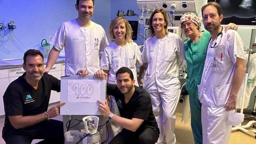
Health in Aragon | Royo Villanova Hospital uses cryotherapy to better detect lung diseases

The Pulmonology Service of the Royo Villanova Hospital in Zaragoza has submitted an application a new method that, thanks to the use of cryotherapy (very low temperatures), makes it possible to better diagnose certain lung diseases such as cancer, tuberculosis or lymphoma. This practice is performed by the newly established team of the bronchoscopy department and is known as cryobronchoscopy of the mediastinum (cryo-EBUS). Since its implementation in November last year About 30 patients have already been successfully tested.“We are very happy,” explains the head of the department, Dr. Javier Lazaro, to this newspaper.
Obtaining a larger tissue sample allows avoiding the surgical alternative. This speeds up the diagnosis in a less invasive way and avoids possible complications of surgical activity.
Any internal examination of the bronchi is carried out through bronchoscopy, but if there are complications, it is necessary to perform echobronchoscopy, which allows for internal ultrasound of the bronchi. It is then that the new technique becomes relevant, because it “much more accurate, takes a larger sample, of higher quality and, in addition, avoids bleeding.” — explains Lazarus.
“Using cryo-Ebus Instead of puncturing tissue, a tube is inserted into the patient’s airway in real time. and a cryoprobe is inserted through it. Using extremely low temperatures, we freeze the tissue sample for about five seconds to extract it,” he explains. It is by obtaining a larger tissue sample surgical alternatives are avoided. This speeds up diagnostics in a less invasive manner and avoids possible complications of surgical procedures.
The quality of the samples obtained makes it possible to use immunohistochemistry and molecular biology techniques, “which allow us to tailor treatments to patients with lung cancer,” says Lazaro.
Moreover, among the advantages of this method is that avoids additional examinations for the patient“The puncture we used before resulted in the needle collecting loose cells, and if we needed to take more samples, we needed more punctures. Now we can extract the tissue with all its structure in such a way that it is of better quality, we avoid additional studies and tests, because more studies can be done with it,” explains Lazaro.
Once a piece of tissue is obtained, it is analyzed using Pathological anatomy service present during the procedure. Each procedure is performed by two pulmonologists together with the medical staff of the department.
The technique involves great progress has been made in identifying various neoplasms such as lung cancer, lymphoma or metastatic diseases, as well as inflammatory diseases such as sarcoidosis or infectious diseases such as tuberculosis. Thanks to the quality of the samples obtained, it is possible to apply methods of immunohistochemistry and molecular biology, “which allow select individual treatment for lung cancer patients,” Lazaro says.
The operation is performed under sedation and on an outpatient basis in the day hospital of pulmonology.
Proof It usually lasts “about half an hour” if there are no complications. “We’ve had cases that lasted more than an hour,” he adds. Action Complete under sedation and outpatient through the day hospital of pulmonology, from where the patient is transferred to the institutions of interventional pulmonology for the development of the methodology and subsequent discharge.
Thusand avoid the hospital stay they may be subjected to if more invasive diagnostic methods are used. Thus, the introduction of this new method means a reduction in diagnostic time and accuracy, accelerating the selection of treatment and its initiation.
