AI differentiates between cancer and normal cells

A team of scientists from Spain has created innovative artificial intelligence (AI) that can differentiate between cancer cells and normal cells, as well as detect the earliest stages of viral infection inside cells. The results, published in the journal Nature Machine Intelligence, open up new possibilities for developing advanced diagnostic methods and disease monitoring strategies.
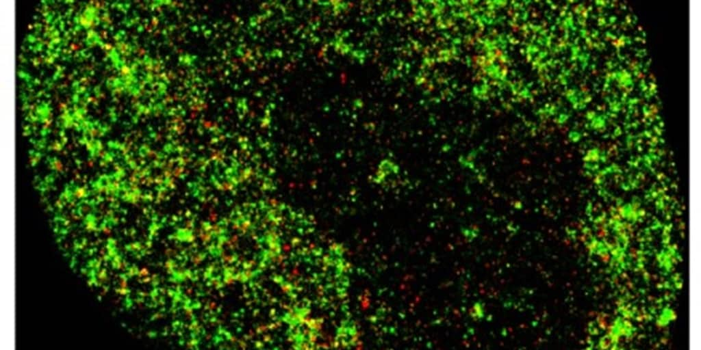
The instrument is called AINU (AI Core) analyzes high-resolution images of cells obtained using an advanced microscopy technique known as STORM. This method produces images with nanometer resolution, revealing detailed structures invisible to traditional microscopes.
The AI is able to detect internal changes in cells as small as 20 nanometers (nm), which is about 5,000 times smaller than the width of a human hair. These changes are so subtle that they are not visible to the human eye using conventional methods. According to ICREA research professor Pia Cosma, co-lead author of the study and a researcher at the Center for Genomic Regulation (CRG) in Barcelona:The resolution of these images is powerful enough that our AI can recognize specific patterns and differences with amazing accuracy.“This includes changes in the organization of DNA inside cells, allowing changes to be detected at very early stages.
AINU is a convolutional neural network, a type of artificial intelligence designed specifically to analyze visual data such as images. In medicine, this type of AI is used to analyze medical images and detect signs of cancer or abnormalities that may go unnoticed by the human eye.
AINU is also capable of detecting viral infections at very early stages. For example, it can detect changes in the nucleus of a cell infected with the herpes simplex virus type 1 just an hour after infection. The AI achieves this by detecting small differences in DNA density that occur when the virus begins to change the cell’s structure.
Ignacio Arganda-Carreras, co-author of the study and Ikerbasca researcher at UPV/EHU, notes that “this method can detect virus-infected cells very soon after infection begins, which could be critical for developing more effective treatments and vaccines.” Additionally, co-author Limei Zhong from Guangdong Provincial People’s Hospital in China adds that “AINU can be used to quickly and accurately diagnose infections from simple blood or tissue samples.”
Despite the promising results, the study authors warn of limitations before the technology can be implemented in a clinical setting. The STORM imaging technique, which is crucial to AINU, requires specialized equipment and can only analyze a few cells at a time, which may not be effective for rapid diagnostics in clinics.
However, the team optimistic about the future. Pia Cosma notes that “advances in STORM imaging technologies could make these microscopes available in small labs and eventually in clinical settings.” In addition, Davide Carnevali, first author of the study and a CRG researcher, emphasizes that AINU could revolutionize stem cell research by speeding up and improving the accuracy of their detection, which would also contribute to reducing the use of animals in science.
While it may take time for AINU to achieve clinical benefits, the tool is already set to accelerate scientific research and open up new possibilities in the diagnosis and treatment of complex diseases.
