Fighting cancer with artificial intelligence

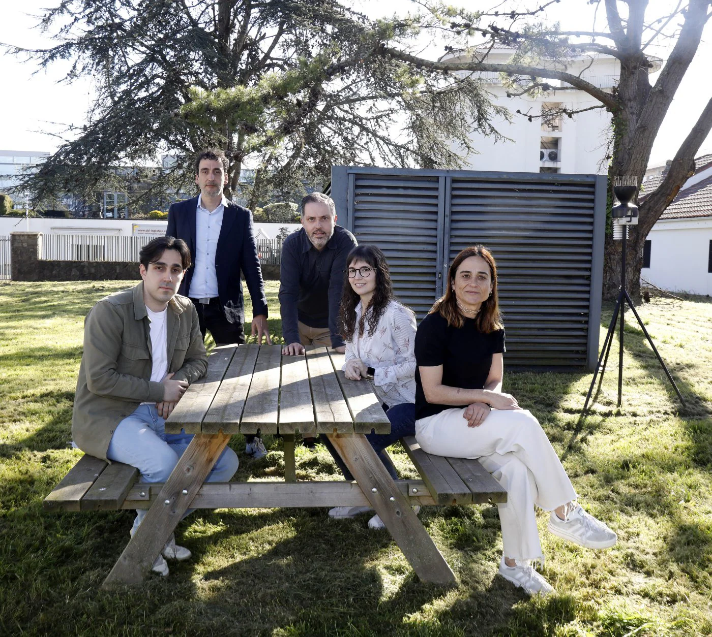
Can artificial intelligence advance cancer research? Maybe. In fact, “we already see its potential.” Its use “will greatly help us personalize the treatment of cancer patients,” says Rene Rodriguez, principal investigator of the sarcoma and experimental therapeutics group at Ispa-Finba. His team is part of the Organaides program, which also includes the Head and Neck Tumor Oncology Research Group led by Monica Alvarez, the Idonial Foundation and Izertis.
Funded by the former Idepa (now transformed into Sekuens), its raison d’être is to “create tumor organoids through 3D printing in order to be able to observe the evolution of tumors using artificial vision techniques,” summarizes Saray Gonzalez, Head of Innovation projects. . in Isertis. The ultimate goal is to predict what the prognosis or most appropriate treatment will be for each case.
Organoids are three-dimensional laboratory replicas of tumors seen in patients. This patient-derived pattern is achieved after a biopsy or surgery to remove the tumor. “Fresh samples come into the laboratory and we process them there to obtain the organoid,” says Rene Rodriguez. This task was carried out by Asturian researchers using traditional methodologies, and later Idonial improved it by introducing 3D bioprinting methods. In this way, it was possible to “systematize” the process, make it “more reproducible” and “reduce time,” explains Manuel Alejandro Fernandez, head of the foundation’s health department.
It is in their laboratories that the three-dimensional environment in which these organelles must develop is bioimprinted. An environment that must be personalized for each tumor type. “We are trying to adapt this material to the needs of organoids. And in addition to working with inks or bioinks, when the composition includes organoids, we work on the design, since it has been documented that the 3D structure you print directly affects the results of the organoids,” says Foundation Idonial researcher Helena Herrada.
When the Organaides project was launched in 2022 and will end at the end of this year, Isertis included a new variable in the effort: artificial intelligence. Based on the microscopic images provided by the Ispa-Finba research groups—in which cells and organoids are visualized—“we developed a model capable of detecting these organoids artificially, without human intervention.”
The AI not only detects them, but is also able to identify “different organelle morphologies,” says Samuel Kamba, an artificial vision expert at Izertis. All this information generates a set of numerical data that can be used to create evolutionary graphs to see the organelle’s growth over time or track its shape. Also develop statistical models or other types of artificial intelligence that will provide more information in the future, such as what parameters may influence organoid size.
“With artificial intelligence, we can have reliable models that can help us anticipate what will happen in the clinic during treatment. This can help us analyze organelles in a way that we could not otherwise achieve, and also in a more systematic and faster way. This can help us obtain parameters of organoid growth and drug response. This should help us correlate the parameters provided by artificial intelligence with the response that the patient actually has. Being able to predict how an organoid will grow, whether it will respond to a certain drug or not… This is a long-term goal,” emphasizes Rene Rodriguez.
This is the main advantage. Thanks to this, without the need for any invasive technique, it is possible to know what can happen in each specific case, depending on the characteristics of the patient and the tumor. All this “with fewer tests” and “more accurate” results, emphasizes Saray Gonzalez, also noting the savings achieved in “time and resources.”