Cubic millimeter of human brain with nanometer resolution
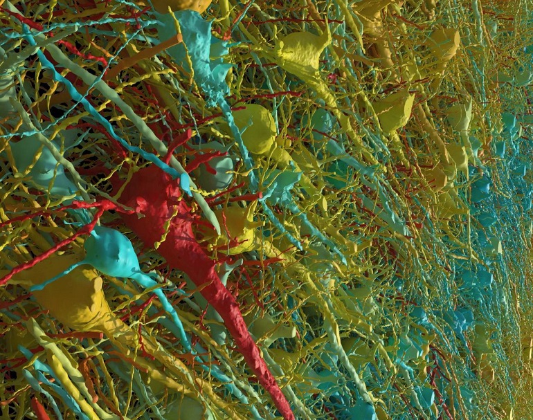
It shows each cell and its network of neural connections in great detail. donated fragment of human temporal cortex which is about half a grain of rice.
Achievement published in the latest issue of the magazine The science, is the latest result of nearly 10 years of collaboration with Google Research scientists. The team combines Lichtman electron microscopy images with artificial intelligence (AI) algorithms for color coding and reconstruct a very complex brain structure mammals.
The goal of this collaboration, funded by the BRAIN Initiative of the US National Institutes of Health, is to create a high-resolution map of the neural connections of the entire mouse brain. This would be about 1,000 times the data they just obtained from a one-cubic-millimeter fragment of human cortex.
“The word ‘fragment’ is ironic,” says Lichtman, the Jeremy R. Knowles Professor of Molecular and Cellular Biology and dean of the natural sciences at Harvard. “For most people, a terabyte is gigantic, but this tiny piece contains 57,000 cells, 230 millimeters of blood vessels and 150 million synapses, which is equivalent to 1,400 terabytes of data.”
Judging by the comments Lichtman at SINC via email, “the purpose of this work is to present Details of the structure of the human brain with enough resolution to see all the synaptic connections between nerve cells.”
Essentially, he adds, “this is an engineering task that requires staining the brain with heavy metals (osmium, lead and uranium), cutting ultrathin sections of the brain (with an automatic ultramicrotome with a diamond blade), obtaining high-speed imaging with an electron microscope multibeam scanning and a range of computational methods for merging, aligning, segmenting and annotating image data.

The study leader emphasizes: “Many of these computational steps required machine learning and artificial intelligence. Our teams at Harvard and Google have worked diligently for several years to achieve this.”
Unseen details of the brain structure
The new map reveals never-before-seen details of the brain’s structure, including a rare but powerful set of axons connected by up to 50 synapses. The team also noticed oddities in the tissue, such as a small number of axons forming extensive whorls. Since the sample was taken from patient with epilepsyResearchers aren’t sure whether these unusual formations are pathological or just weird.
The Lichtman deposit is connectomics, which, similar to genomics, aims to create complete catalogs of brain structure, down to individual cells and connections. These complete maps will open the way to new knowledge about brain function and diseaseabout which scientists still know very little.
Google’s advanced artificial intelligence algorithms have made it possible to reconstruct and map brain tissue in three dimensions. The authors also developed a set public access tools which other scientists can use to explore and annotate the connectome.
“Given the huge investment that has gone into this project, which is not reported, it was important to present open resultsso that everyone can benefit from them,” he says. Viren JainGoogle Research researcher and co-author of the paper.
In this sense, Lichtman emphasizes that “the data set is so large that one laboratory will not be able to study it in its entirety. Relevant discoveries are more likely to be made if all interested parties have access to this information.”
On the other hand, the neuroscientist tells SINC that in the reconstruction now presented, they learned that “the human brain as a physical entity is much more complex than the thoughts it produces. This means that understanding our brain through thinking will be quite a challenge.”

In addition, he emphasizes, “we found many beautiful and strange things In this great exhibition they are unexpected and in many cases quite mysterious, some of them we have highlighted in the study and others can be found in the gallery of the page dedicated to the works hosted on Google.
When asked how this detailed reconstruction could lead to a better understanding of brain diseases and the development of new treatments, the neuroscientist replied: “The main hope is that psychiatric diseases and diseases related to brain development have underlying physical pathology “And that our study will help researchers better describe what happens in the brain that causes thought disorders in affected people.”
The next stage of the investigation will be the examination of the case. mouse hippocampus formationimportant to neurobiology due to its role in memory and neurological diseases.
Influence of Santiago Ramon y Cajal
Jeff Lichtman holds the title of Santiago Ramon y Cajal Professor of Arts and Sciences at Harvard. He emphasizes that the Spanish Nobel laureate had a “tremendous influence” on his research: “Cajal was first scientist to specialize in connectomicsdemonstrated through Golgi spotthat neurons are connected in a directed network.”
Lichtman concludes: “Modern connectome work like our study overcomes the only limitation of Golgi staining: dispersed staining. “Now we essentially have a Golgi technocolor, so all the cells and connections are visible in the same pattern.”
Link:
Alexander Chapson-Coe et al. Petavoxel fragment of human cerebral cortex reconstructed at nanoscale resolution. The science2024